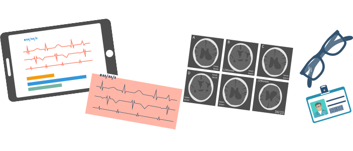
Positron emission tomography (PET) scans are used to produce detailed three-dimensional images of the inside of the body.
It is a nuclear medicine functional imaging technique that is used to observe metabolic processes in the body. The system detects positron-emitting radionuclide (tracer), of which the radiographer will inject a very small dose into the body. 3-D images of tracer concentration within the body are then constructed by computer.
PET scan can also sometimes tell if an area in the body is scar tissue or an active cancer. The concentrations of tracer imaged will indicate tissue metabolic activity enabling the tracer to reveal the possibility of cancer metastasis (i.e., spreading to other sites).